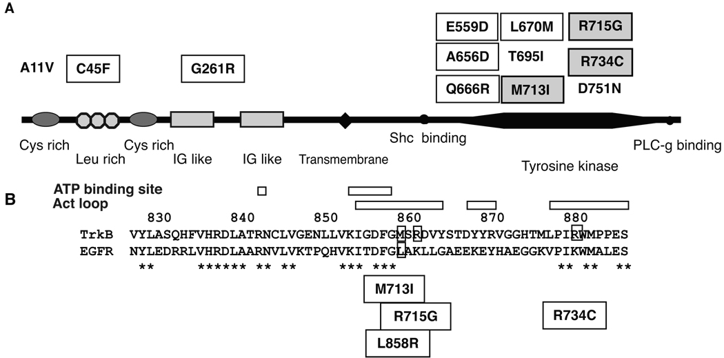
Figure 1.
(A) Schematic representation of TrkB domain, Cys rich, cycteine rich region; Leu rich, leucine rich region; IG like, immunoglobulin like-domain. Reported non-synonymous mutations are indicated. Mutations with square are reported in lung adenocarcinoma (15). Mutations with shaded square are reported in LCNEC (16). A11V was found in glioblastoma (36). T696I and D751N are found in colon cancer (37). (B) Protein alignment of TrkB and EGFR. * indicates matched amino acid sequence. Positions of activation loop and ATP binding site are indicated. Positions of EGFR mutation L858R and TrkB mutations, M713I, R715G and R734G, are also indicated.

