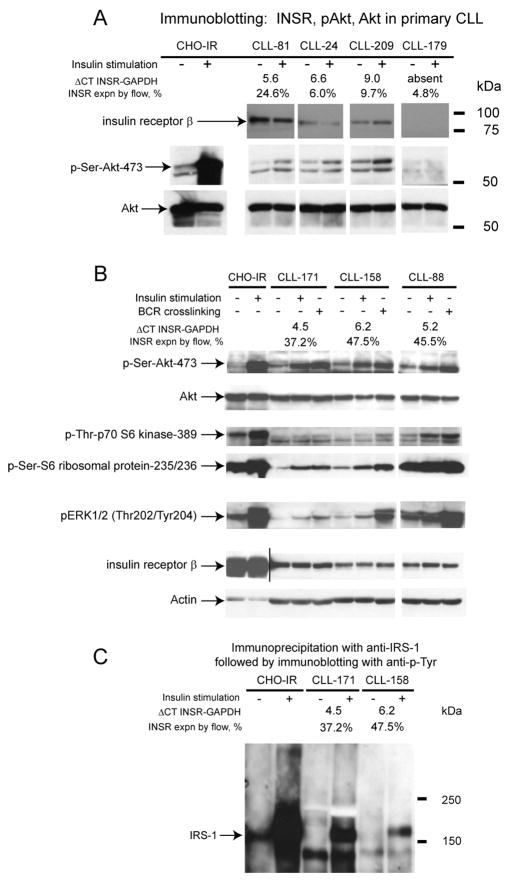
Figure 4. The INSR activates canonical INSR signal transduction pathways in CLL.
Panel A-B: CD19+ cells from selected CLL cases were cultured and left untreated or stimulated with 10nM insulin. Detergent lysates were prepared, protein was fractioned by SDS-PAGE, transferred to membrane and subsequently prepared for immunoblotting with various antibodies as indicated. For data presented in panel B, aliquots of cells were also stimulated in parallel with a combination of biotinylated anti-IgM F(ab)2 fragments together with avidin. C: Immunoprecipitation of IRS1 out of CHO-IR or CD19+ CLL-derived cells left untreated or treated with 10nM insulin followed by immunoblotting with anti-phosphotyrosine antibodies. The approximate position of pre-stained molecular weight markers is indicated.

