
Figure 2.
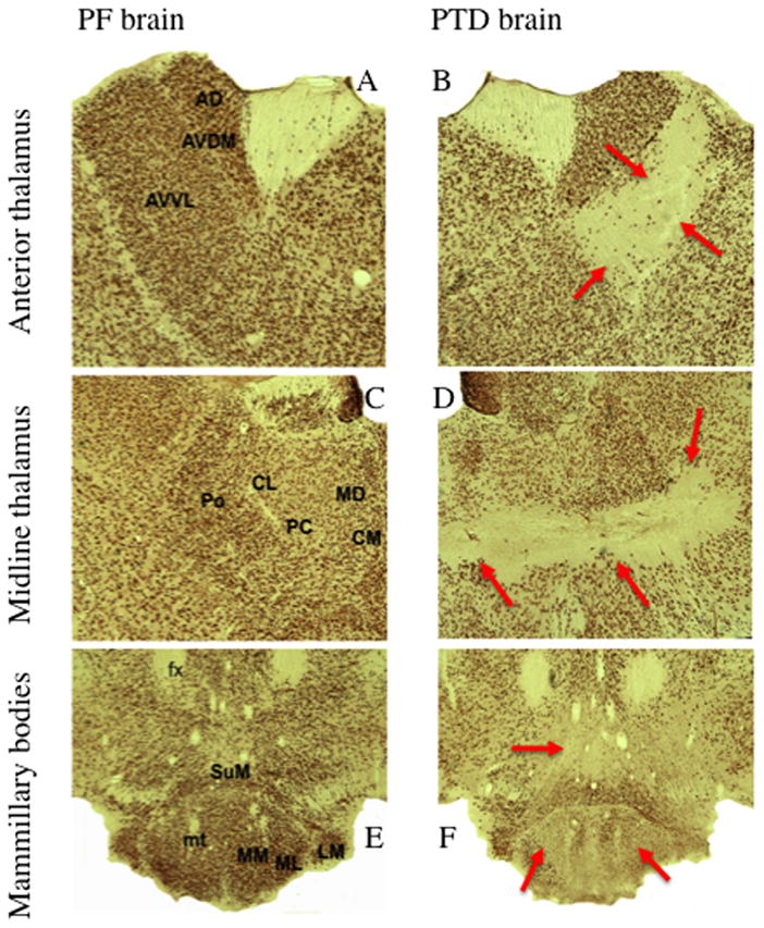
Neuronal specific nuclear protein (NeuN) stained slides comparing the thalamus and mammillary bodies of PF and PTD rats (from Anzalone et al, 2010). The top row (A and B) reveal the anterior thalamic nuclei in both PF and PTD rats. In PTD rats there is selective cell loss in the anteroventral ventrolateral (AVVL) with relative sparing of the anteroventral dorsal medial (AVDM) and anterodorsal nuclei (AD). The middle row (C and D) are images of midline and intralaminar thalamic structures. There is significant cell loss in the intralaminar nuclei (central medial (CM), paracentral (PC) and centrolateral (CL) nuclei) as well as the posterior thalamic nucleus (Po). However, the medial dorsal (MD) nucleus is spared—with the exception of the most ventral tip. The bottom row (E and F) are examples of the hypothalamus including the mammillothalamic tract (mt), medial mammillary nucleus (MM), lateral medial mammillary nucleus (ML) lateral mammillary nucleus (LM) and the supramammillary nucleus (SuM). There is significant cell loss medial mammillary ncuclei (MM, ML) and within the SuM.