Figure 7.
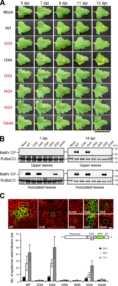
TGBp3 sorting mutants are defective in viral cell-to-cell movement. (A) Leaves of C. quinoa inoculated with various BaMV viruses were photographed on the days post-inoculation (dpi) as indicated. The critical residues for TGBp3 localization are marked in red. Bar, 5 cm. (B) Various viruses were used to inoculate N. benthamiana plants. The inoculated and upper non-inoculated leaves were harvested and lysed at 7 and 14 dpi. Samples were subjected to immunoblot analysis with anti-BaMV CP antibody. Coomassie blue staining for the plant RuBisCO protein served as a loading control. (C) Various BaMV-GFP viruses harboring the TGBp3 mutants indicated were used to inoculate C. quinoa. At 24, 48, and 72 h after inoculation, at least 15 foci on each inoculated leaf were imaged by confocal microscopy. Images show foci that were selected by random at 48 h after inoculation with mock or the indicated viruses. The number of epidermal cells in each infection focus were counted, and data are plotted as mean ± SD (error bars). The scheme of BaMV-GFP reporter virus is depicted above the plot. The red fluorescence was from autofluorescence of plant chloroplasts. Bar, 50 µm.

