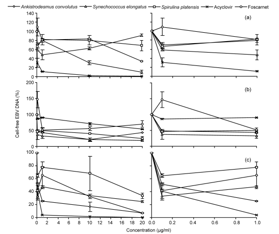
Fig. 2.
Percentage of cell-free EBV DNA in the media of Akata (a), B95-8 (b), and P3HR-1 (c) cells treated with methanol extracts from microalgae or antiviral drugs, compared to the control
The Burkitt’s lymphoma (BL) cells were chemically induced into the lytic cycle before being exposed to microalgal extracts or antiviral drugs for 72 h. DNA was extracted from the culture medium and BamH1-W LP fragment region of the EBV genome was quantified using real time-PCR. The data are shown as mean±SD of two independent experiments performed in duplicate

