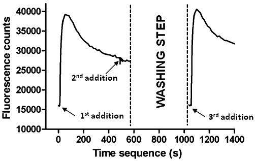
Figure 2.

The figure shows the activation, desensitization and recovery from desensitization of human α6/3β2βV273S-nAChRs stably expressed in HEK293 cells, as tested in the FLIPR Ca2+ influx assay. Cells were loaded with the fluorescent Ca2+ indicator dye FLUO-4-AM and transferred to the FLIPR platform for the measurement of increases in intracellular Ca2+. Increases in the relative fluorescence units represent increases in intracellular Ca2+. Following the first addition of 200 nM nicotine, there was a peak of fluorescence change, reflecting channel activation and slow desensitization. Addition of the same quantity of nicotine to the same cells (second addition) did not yield an effect, confirming receptor desensitization. When cells plate was extensively washed with the assay buffer, residual fluorescence disappeared and addition of 200 nM nicotine (third addition) caused a further fluorescence increase, revealing recovery from desensitization. Data are from a representative experiment repeated at least three times with similar results.
