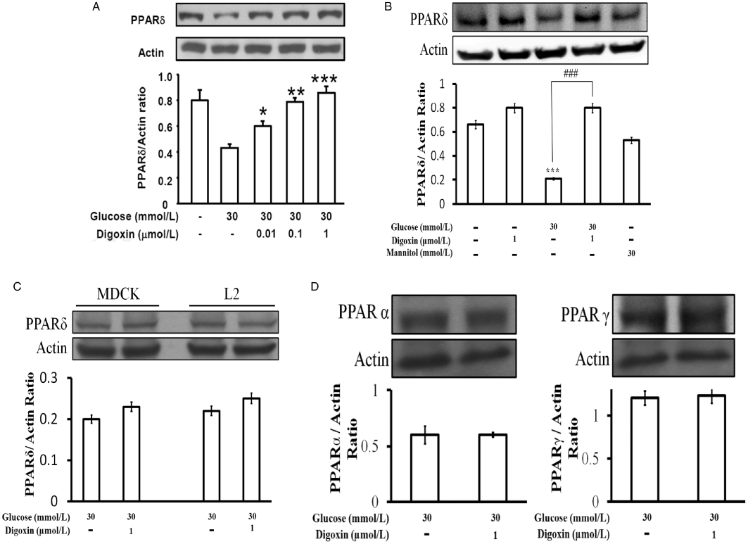
Figure 1.

Effects of digoxin on PPARδ expression in H9c2 cells. (A) H9c2 cells were cultured with or without glucose at 30 mmol·L−1 and treated with digoxin, which was also incubated with 5.5 mmol·L−1 glucose. (B) Cells were also exposed to 24.5 mmol·L−1 mannitol to produce the same osmolarity (317 mOsmol·L−1) as that produced with the highest concentration of glucose (30 mmol·L−1). The embryonic rat H9c2 cells (A), rat lung L2 cells and MDCK cells (C) were cultured with 30 mmol·L−1 glucose for 24 h, and then treated with digoxin (0.01–1 µmol·L−1) for 30 min. Also, the possible effects of digoxin (1 µmol·L−1) on the expressions of PPARα and PPARγ in H9c2 cells were also investigated (D). These cells were harvested to determine the protein levels by Western blot analysis. All values are presented as mean ± SEM (n = 4 per group). *P < 0.05, **P < 0.01 and ***P < 0.001 as compared with the HG-treated cells.
