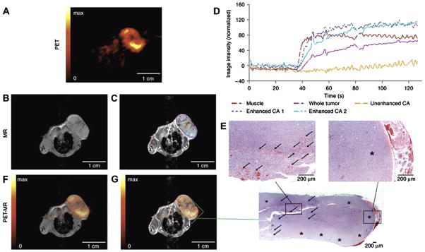
Fig. 8.
Fully integrated dual modality PET/MR imaging of a subcutaneous tumor. (A) PET images of (18F)FLT tracer uptake indicate regions of high cell proliferation (yellow) and necrosis (black). (B and C) Pre- and post-contrast MR images obtained using the same device reveal the underlying anatomy and further define sites of tissue necrosis (C, yellow region of interest in center of tumor). (D) Dynamic measurement of contrast enhancement within regions of interest highlighted in (C). (F and G) Coregistration of PET and MR images reveals areas of inflammation and necrosis that were confirmed by histology (E). Note that integration of the hardware for PET and MRI permits direct image coregistration as the anatomy of experimental subject was not shifted; as is often the case when moving patients between two imaging devices. Reproduced, with permission, from Ref. [14].

