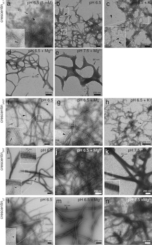
Fig 6.

Ultrastructure of negatively stained crescentin mutants. Samples of purified crescentin mutants (0.2 mg/mL, ∼ 4 μM) are in 50 mM PIPES (pH 6.5), HEPES (pH 7.5), or Tris-HCl (pH 8.5) unless otherwise noted. K+ and Mg2+ denote 200 mM KCl and 5 mM MgCl2, respectively. (a) CrescentinΔL1 in 5 mM buffer. Single 9-nm-wide filaments are denoted with arrows, and filaments with approximate widths of 18 nm (arrowhead) or 27 nm (asterisk) are marked. (b) CrescentinΔL1 in 50 mM buffer. The arrowhead denotes an 11 nm-wide filament. (c) CrescentinΔL1 in the presence of 200 mM K+. Arrowheads mark globular structures with approximate diameters of 20–30 nm. (d) CrescentinΔL1 in the presence of 5 mM Mg2+. The arrow marks a 50 nm-wide structure. (e) CrescentinΔL1 at pH 7.5 with 5 mM Mg2+. A structure with 80 nm width is marked (arrow). (f) CrescentinΔN27 in 50 mM buffer. Arrows denote 8–9 nm-wide filaments (also in inset), the arrowhead denotes a 16 nm-wide filament, and the asterisk denotes a 24 nm-wide filament. (g) CrescentinΔN27 in the presence of 5 mM Mg2+. Arrow in inset denotes a 9 nm-wide filament, and the arrowhead denotes a 17 nm-wide filament. (h) CrescentinΔN27 in the presence of 200 mM K+. Arrow denotes a 22 nm-wide filament. (i) CrescentinSAT. A single 9 nm-wide filament (inset, arrowhead) and 40 nm-wide bundles (arrows) are denoted. Bundled areas showed an axial repeat of 57.1 ± 0.7 nm (n = 25). (j) CrescentinSAT in the presence of 5 mM Mg2+. The arrowhead marks a 10 nm-wide filament. (k) CrescentinSAT at pH 7.5 in the presence of 5 mM Mg2+. The paracrystalline structures have variable width and an axial repeat of 58.7 ± 0.3 nm (n = 30). Inset shows a detail of the striations. (l) CrescentinΔT. Single filaments (arrowheads, inset) displayed widths of 17–20 nm. (m) CrescentinΔT in the presence of 5 mM Mg2+. Tapered paracrystalline bundles had an axial repeat of 55.6 ± 0.6 nm (n = 30). (n) CrescentinΔT at pH 7.5 in the presence of 5 mM Mg2+.
