
Figure 2.
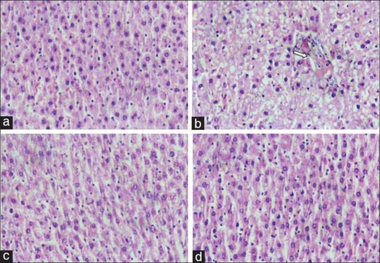
(a) Normal architecture of rat hepatic lobule (×40) H & E stained. (b) Fatty liver tissue with marked diffuse granular degeneration and mild multifocal biliary hyperplasia (×40). (c) Histology of liver tissue treated with standard silymarin showing normal architecture (×40) H & E stained. (d) Histology of liver tissue treated with P. kurroa 400 mg/kg showing normal architecture (×40) H & E stained.