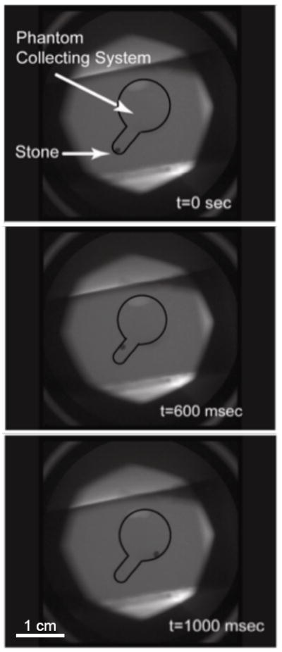
Fig. 4.

Fluoroscopy monitoring of an artificial stone that was repositioned from the lower pole into the collection system of a kidney phantom. Displacement of the stone was seen immediately after the application of focused ultrasound and the total distance traveled was approximately 1 cm. Estimated velocity magnitude was 1 cm/s.
