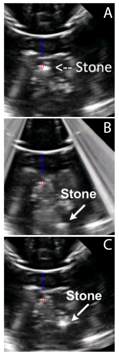
Fig. 5.

Ultrasound monitoring of an artificial stone (a) before, (b) during, and (c) after delivering focused ultrasound to move the stone from the lower pole to the collecting system of the kidney phantom. Blue artifacts were added to denote the axis of the focused array, and the red artifact shows its focus; (a) shows initial targeting of the stone. The lower pole appears at the top of these images because the hand-held device was in contact with the bottom of the phantom.
