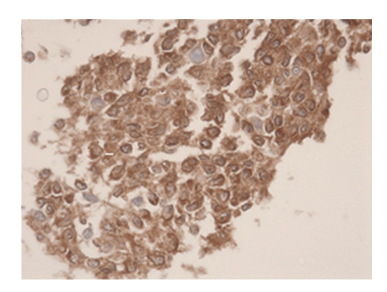
Figure 3.

Immunohistochemical stain CD79A (HE 400×). Microscopical report: histological tissue fragments are presented. The image is determined by small lymphatic cells with irregular shaped nucleoli. The chromatine-pattern is enlarged. There is indentation on multiple sides. Some B-lymfoblasts are seen (CD79A/20). The total image is representative of a follicular non-Hodgkin lymphoma.
