
Fig. 1.
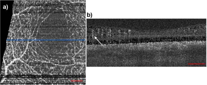
The human parafoveal capillary network, extracted using phase variance analysis with swept-source OCT at 1050 nm. (a) Fundus projection taken from the inner retinal layers. (b) Phase variance tomogram across the blue line in (a). The arrow indicates typical vessel shadow decorrelation artifacts. The red bars denote 200µm.