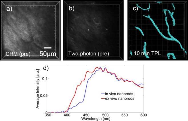
Fig. 3.

Representative two-photon microscopy images of intravenously delivered GNRs in hamster model with accompanying confocal reflectance image (a) Confocal reflectance image of tissue showing location of blood vessels which appear dark against the surrounding tissue (b) High power (20 mW) two photon image of the same vascular region prior to intravenous injection of GNRs (c) Two-photon image using low incident power of 1 mW following intravenous injection GNRs showing blood vessels in the tissue. Two-photon microscopy of vascular sites prior to GNR injection, using 1-20 mW incident power, yielded no detectable signal from blood vessels. Asterisk denotes the same vessel junction on all three images displayed. (d) Spectral profile of GNRs in vitro and within in vivo blood vessels following intravenous injection.
