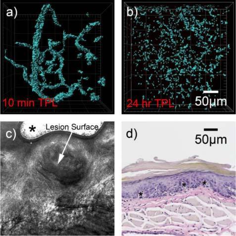
Fig. 4.

(a) Two-photon 3D reconstructed image of precancerous (dysplastic) lesion labeled with GNRs 10 minutes post-inoculation, showing dense and tortuous network of blood vessels, obtained with an incident power of 1 mW. (b)

(a) Two-photon 3D reconstructed image of precancerous (dysplastic) lesion labeled with GNRs 10 minutes post-inoculation, showing dense and tortuous network of blood vessels, obtained with an incident power of 1 mW. (b)