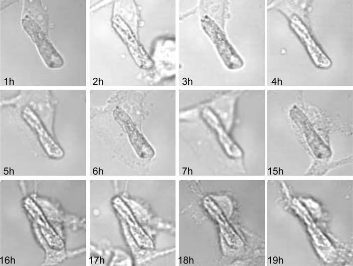
Fig. 6.

Evolution of a fibroblast culture as observed by phase contrast microscopy. The chosen zone corresponds to a dust artefact around which cells are transforming. Sequence of images after digital registration by the PRM method. Image size: 120 × 120 pixels; 48 × 48 μm2; 60 × oil lens N.A.=1.42.
