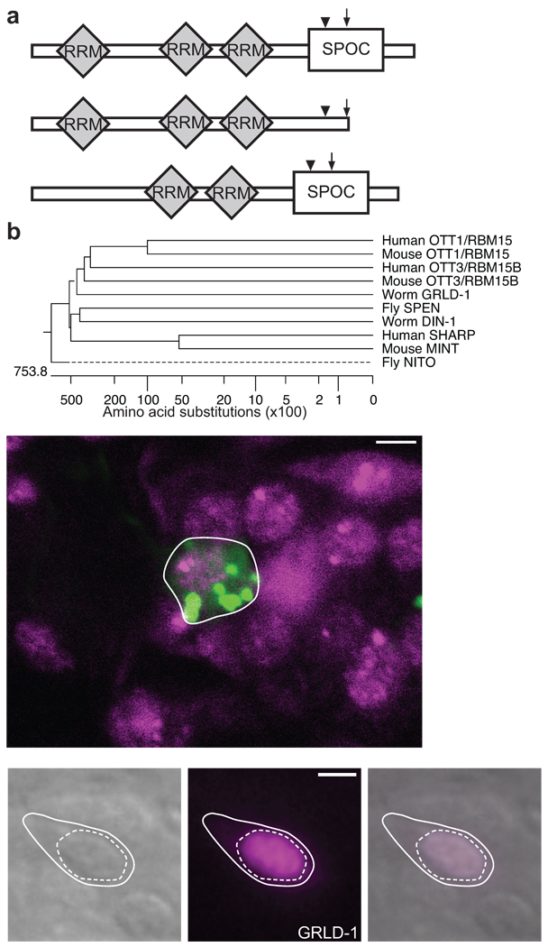
Figure 3. GRLD-1 is a member of the SPEN family.
(a) Schematic domain structure of the three isoforms of GRLD-1. The arrow indicates the position of the molecular lesion of grld-1(wy225) and the arrowhead indicates that of grld-1(wy655). (b) Phylogenetic analysis of GRLD-1 and SPEN family members. (c) grld-1 is expressed in AVE. mCherry (pseudo-colored green) is expressed in AVE by the opt-3 promoter (outlined by white line) and GFP-tagged GRLD-1 (pseudo-colored magenta) is expressed with fosmid recombineering. The nerve ring is anterior to AVE. The image is a single confocal plane (~1 µm) of an L2-staged worm. Scale bar, 2 µm. (d–f) GFP::GRLD-1 localizes to the nucleus when expressed in AVE. Solid line, cell body; dashed line, nucleus. DIC image of AVE (d), pseudo-colored GFP-tagged GRLD-1 (e), and overlay (f) at the L4 stage. Scale bar, 2 µm.

