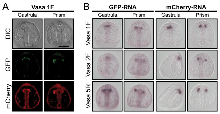
Figure 1. Small micromere retention of microinjected synthetic RNA.
(A) GFP (green) fluorescence from a Vasa- GFP mRNA construct assayed for protein expression in gastrula and prism-stage embryos along with mCherry (red) control mRNA. (B) In situ RNA hybridization for GFP or mCherry synthetic mRNA in gastrula and prism-stage embryos. Vasa 1F is a full-length ORF fused to Xenopus β-globin 5′ and 3′ UTRs. Vasa 2F-GFP is the same construct lacking the Vasa region N-terminal to the Zn-fingers, and Vasa 5R-GFP RNA instead lacks the catalytic domain and the helicase domain. GFP and mCherry RNA probes detect the spatial distribution of the exogenous RNA. Note that the Vasa protein accumulates selectively in the small micromeres on the left of the embryo, whereas the mRNA is retained equally in the small micromeres of both sides. Scale bar = 50 μm.

