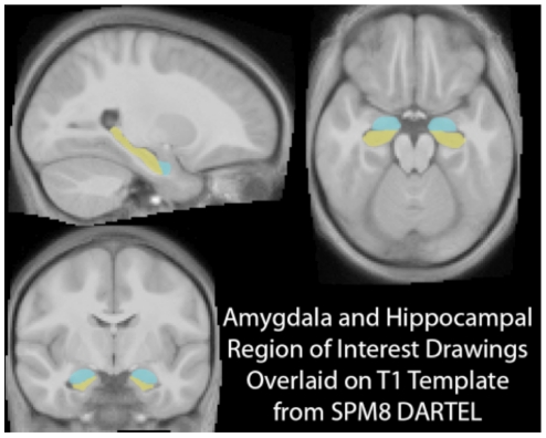
Figure 1. Hippocampal and amygdala region of interest drawings.
The top left brain slice shows a sagittal brain slice with the hippocampus highlighted in yellow and the amygdala in turquoise, while the top right brain image shows an axial slice (with the hippocampus again highlighted in yellow and the amygdala in turquoise). The bottom left brain picture shows a coronal slice with the amygdala in turquoise and the hippocampus in yellow.

