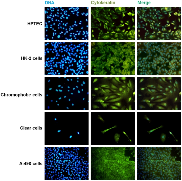
Figure 4. Fluorescence microscopy images from immunocytochemical staining for cytokeratin.
Hoechst 33258, anti-cytokeratin antibody staining, and merged images with double staining of primary cultured HPTEC, clear cell and chromophobe RCC cells. HK-2 and A-498 cells were used as control of human normal and tumor epithelial kidney cells, respectively. Magnification: 200x.

