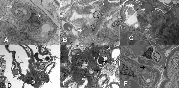
Figure 3.
Electron microscopy showing (A) double contours with subendothelial deposits and new basement membranes in case 1; (B) mesangial deposits in case 2; (C) subepithelial, subendothelial, and mesangial deposits in case 3; (D) mesangial, subepithelial, and subendothelial deposits in case 4; and (E and F) osmiophilic wavy sausage-shaped intramembranous deposits and mesangial deposits in cases 4 and 5, respectively (white arrows, electron dense deposits; black arrows, dense deposits along basement membrane).

