
Figure 3. The interactions between RXR and VDR when bound to DNA.
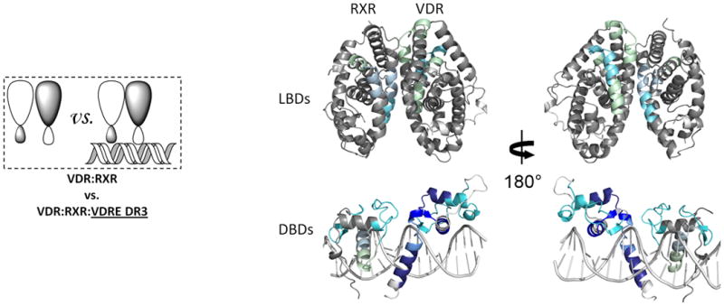
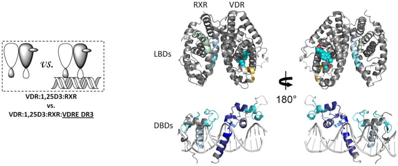
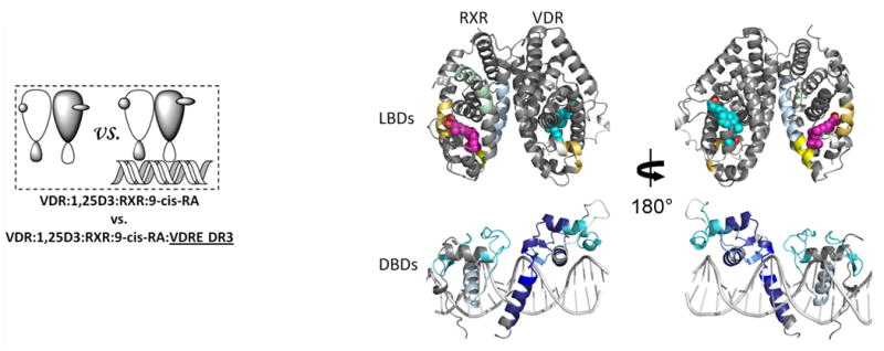
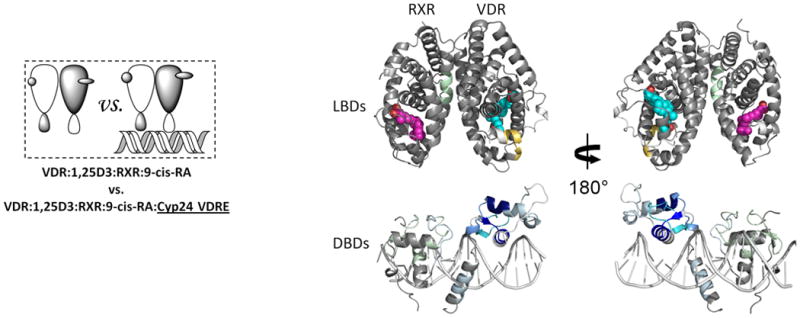
Differential HDX data mapped onto RXR-VDR docking model of DBD and LBD domains when bound to different ligands and DNA response elements. (a) The interactions between RXR and VDR on VDREdr3 in absence of ligands (Supplementary Tables 1b(vi) and 1c(vi)). (b) The interactions between RXR and VDR on VDREdr3 in presence of 1,25D3 only (Supplementary Tables 1b(vii) and 1c(vii)). (c) The interactions between RXR and VDR on VDREdr3 in presence of 1,25D3 and 9-cis-RA (Supplementary Tables 1b(viii) and 1c(viii)). (d) The interactions between RXR and VDR on Cyp24vdre in presence of 1,25D3 and 9-cis-RA (Supplementary Tables 1b(ix) and 1c(ix)).