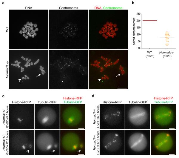
Figure 7. Reduced numbers of chiasmata form in Hormad1−/− oocytes.
a, Centromeres were detected by IF and DNA was detected by propidium iodide on nuclear spreads of in vitro-matured metaphase stage oocytes. In WT cells, 20 pairs of chromosomes are connected by chiasmata. In the Hormad1−/− mutant, a large fraction of chromosomes does not have chiasmata (one such chromosome is marked by arrowhead). Arrow marks a pair of chromosomes connected via a chiasma in the mutant oocyte. Note that bivalents are symmetrical and chiasmata form between chromosomes of identical length in the mutant, indicating that CO formation took place between homologous chromosomes. Chromosomes scatter over a larger area during nuclear spreading in the mutant, therefore it was not possible to include all 40 chromosomes of a meiosis I oocyte in the image. Bars, 10 μm. b, The average numbers of paired chromosomes connected by chiasmata (marked by line) are reduced nearly three-fold in in vitro-matured Hormad1−/− oocytes relative to WT oocytes. c and d, mRNAs encoding β-tubulin-GFP and histone H2B-RFP were injected into WT (c) and Hormad1−/− (d) oocytes during the germinal vesicle stage (prophase), and the oocytes were matured in vitro. The fluorescent proteins in the oocytes were imaged 5.5 hours after germinal vesicle break down (GVBD), at a time when WT oocytes are in the first meiotic metaphase, and 17.5 hours after GVBD, at a time when WT oocytes are arrested in the second meiotic metaphase. A polar body (marked by arrowhead in c), which was extruded 7 hours after GVBD (Supplementary Information, Movie 1) is observed next to the metaphase II stage WT oocyte at the 17.5 hour time point. In the Hormad1−/− oocyte (d), the meiotic spindle is abnormally long and chromosomes fail to align at both time points. Meiotic anaphase did not take place in the displayed Hormad1−/− oocyte (Supplementary Information, Movie 2), and no polar body can be observed at either time points (d). Bars, 20 μm.

