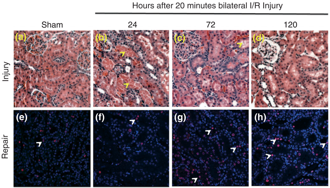
FIGURE 1.
Kidney tissue injury (a–d) and repair (e–h) over time following 20 min of bilateral renal ischemia/reperfusion injury. Male Wistar rats were subjected to sham or bilateral ischemia by clamping the renal pedicles for 20 min and then removing the clamps and confirming reperfusion. Rats were euthanized at various times and kidney tissues were collected. Representative photomicrographs of H&E-stained paraffin-embedded kidney sections (at 200 × magnification) and immunohistochemistry for Ki67 (at 400 × magnification) are presented from the following time points: (a, e) Sham surgery; (b, f) 24 h; (c, g) 72 h; and (d, h) 120 h. All fields were chosen from the cortex and outer medulla. Arrows in panels b and c indicate sloughing of cells, tubular dilation and necrosis. Arrows in panels e–h show Ki67 positive nuclei as an indicator of tubular epithelial cell proliferation.

