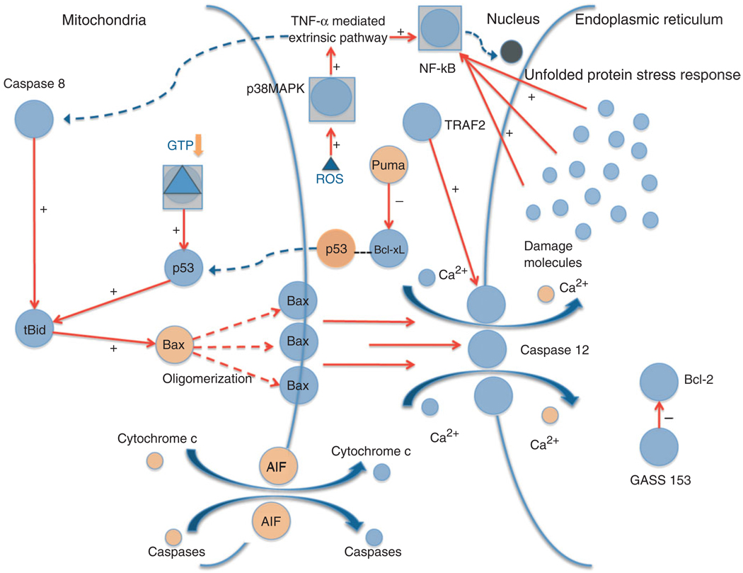
FIGURE 3.
Apoptosis: intrinsic, extrinsic, and endoplasmic reticulum (ER) stress mechanisms. The diagram shows the various signaling pathways leading to apoptosis and the generation of ROS. Upon stress activation, p53 translocates to the mitochondria where it interacts with the BcL-2 family members, including anti-apoptotic BcL-xL and pro-apoptotic Bax and Bak. P53 disrupts inhibitory complexes formed with tBid and Bim, which effectively activate Bax and Bak to oligomerize and form lipid pores in the outer mitochondrial membrane, a process called mitochondrial outer membrane permeabilization (MOMP). The Bax/Bak pathway subsequently activates caspase 12 mediated apoptosis through the release of Ca2+ into the cytoplasm. Cytoplasmic p53 also plays a role in transcription independent apoptosis by being liberated from BcL-xL by Puma, and then activating cytosolic Bax. TNF-α is responsible for extrinsic death receptor mediated apoptosis, which is proposed to be activated by ROS stimulation through the p38 MAPK pathway. This leads to NF-κB and caspase 8 activation, which link the extrinsic pathway to the intrinsic apoptotic mechanism initiated by p53. The ER stress response mechanism might be triggered by the accumulation of ‘damage molecules’ in the ER, stimulating the unfolded protein response (UPR) and activating NF-κB. GADD-153 activates pro-apoptotic factors and downregulates anti-apoptotic BcL-2. Tumor necrosis factor receptor-associated factor-2 (TRAF-2) and caspase 12 stimulation contribute to apoptosis and cell death.

