
Fig. 1.
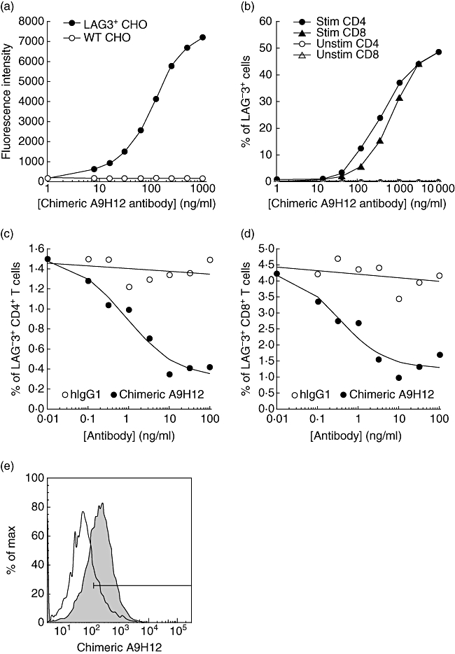
In vitro characterization of the chimeric A9H12 monoclonal antibody (mAb). Binding of chimeric A9H12 mAb at indicated concentrations to control or lymphocyte-activation gene-3 (LAG-3) Chinese hamster ovary (CHO) cells (a) and to Staphylococcal enterotoxin B (SEB)-stimulated (Stim) or resting (Unstim) peripheral blood mononuclear cells (PBMCs) (b). A fluorescein isothiocyanate (FITC)-conjugated anti-human immunoglobulin (Ig)G was used to reveal the chimeric A9H12 mAb bound to the cells. The results in (a) represent the median of fluorescence intensity as a function of antibody concentrations. The graph in (b) presents the percentage of LAG-3+ cells in CD4+ and CD8+ T cell subpopulations. (c,d) An antibody-dependent cell cytotoxicity (ADCC) assay was performed with PBMCs from a cytomegalovirus (CMV)-positive human donor stimulated with a CMV peptide pool for 5 days. Various concentrations of chimeric A9H12 mAb or human IgG1 were added for 4 h and the viability of activated CD4+ or CD8+ T cells was assessed by flow cytometry. Data shown are from one representative experiment of five. (e) Cross-reactivity of chimeric A9H12 with the baboon was assessed by flow cytometry. Target cells were concanavalin A-activated (shaded profiles) or resting (empty profiles) baboon PBMC.