
Fig. 2.
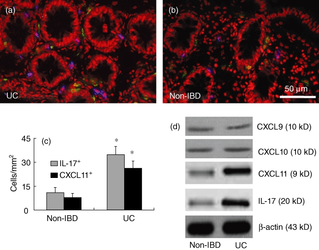
CXCL11 is correlated with the increase in interleukin (IL)-17 in inflammatory bowel disease (IBD) patients. Colonic biopsies were obtained from the 16 ulcerative colitis (UC) patients (a) as described in the text. Six non-IBD samples (b) were obtained from the marginal tissue of surgically removed colonic cancer (proved by a pathologist). (a–c) Cryosections were prepared with the colonic samples and stained with anti-CXCL11 and anti-IL-17 antibodies. The sections were observed under a confocal microscope. The representative confocal images show CXCL11+ cells (in blue) and IL-17+ cells (in green) in colonic tissue. The positive cell counts were presented in (c) as cells per mm2 (mean ± standard deviation; averaged from 20 fields of each subject). *P < 0·05, compared with non-IBD group. (d) A piece of colonic tissue from biopsies [as described in (a–c)] was processed for Western blotting to determine the protein levels of CXCL9, CXCL10, CXCL11 and IL-17. The representative immune blots show the levels of CXCL9, CXCL10, CXCL11 and IL-17 in colonic tissue of non-IBD subjects and UC patients. β-actin was used as internal control.