
Fig. 3.
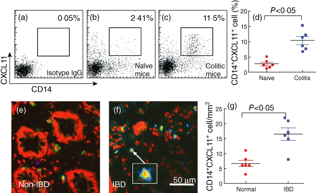
CD14+ cells express CXCL11. (a–d) A trinitrobenzene sulphonic acid (TNBS) colitic mouse model was developed. Lamina propria mononuclear cells (LPMCs) were isolated from mouse colon, stained with anti-CXCL11 and anti-CD14 antibodies and analysed by flow cytometry. The flow cytometry dot-plots show frequency of CD14+ CXCL11+ cells (the gated cells). (d) Summarized data of flow cytometry (each dot represents an individual datum). (e–g) Inflammatory bowel disease (IBD) patients' colonic biopsy tissue was processed for immunohistochemistry to examine the CD14+ CXCL11+ cells. The representative confocal images show CD14+ cells (in blue), CXCL11+ cells (in green) or CD14+ CXCL11+ cells (cells were stained in both green and blue or light blue; see the insert). (g) The immune-positive cell counts (each dot represents a datum from one sample that was averaged from 20 high-power fields, × 200).