Figure 5.
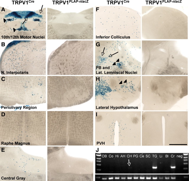
Developmental expression of TRPV1 in the brain. Left, lacZ staining in 80 μm sections of TRPV1Cre/R26R-lacZ mice. Right, Lack of nlacZ staining in the same areas in TRPV1PLAP-nlacZ mice, likely attributable to developmental expression that is extinguished in the adult. A–I, Regions include the 10th and 12th motor nuclei of the medulla (A) (arrowheads indicate cell body staining in 10th and 12th motor nuclei; arrows indicate axonal lacZ label in the area postrema and nucleus of the solitary tract), nucleus interpolaris (B), perivolivary region (C), raphe magnus (D), central gray region (E), inferior colliculus (F), parabrachial nucleus (PB; arrowheads) and nucleus of the lateral lemniscus (arrows) (G), lateral hypothalamic nucleus (H, arrowheads), and paraventricular hypothalamic nucleus (PVH; I). J, RT-PCR analysis of brain regions and peripheral tissues to detect presence of TRPV1 mRNA. Note the faint band in caudal hypothalamus lane (arrow). OB, Olfactory bulb; Co, cortex; Hi, hippocampus; AH, anterior hypothalamus; CH, caudal hypothalamus; PG, periaqueductal gray; Ce, cerebellum; Li, liver; Bl, bladder; Cr, cremaster; neg, no cDNA. Scale bar, 500 μm.

