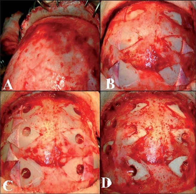
Figure 5.

Intraop pictures showing multiple burr holes drilled over the exposed areas of bone, through small incisions in the perisoteum a) scalp elevation, b) periosteal elevation, c) burr holes and dural opening and d) periosteal flap placed in direct contact with the brain.
