
Figure 1C.
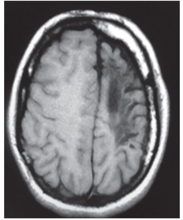
Axial T1-weighted image: Left cerebral hemiatrophy with calvarial thickening. Hypointensity in white matter represents gliosis
Official websites use .gov
A
.gov website belongs to an official
government organization in the United States.
Secure .gov websites use HTTPS
A lock (
) or https:// means you've safely
connected to the .gov website. Share sensitive
information only on official, secure websites.
Axial T1-weighted image: Left cerebral hemiatrophy with calvarial thickening. Hypointensity in white matter represents gliosis