Figure 2.

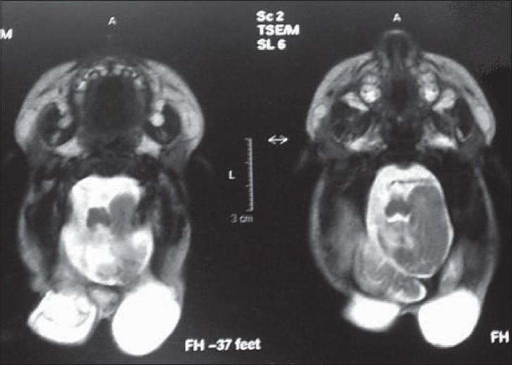
MRI image showing two separate sacs without any neural tissue in them. Note the normal piece of bone in between the two sacs (arrow)

MRI image showing two separate sacs without any neural tissue in them. Note the normal piece of bone in between the two sacs (arrow)