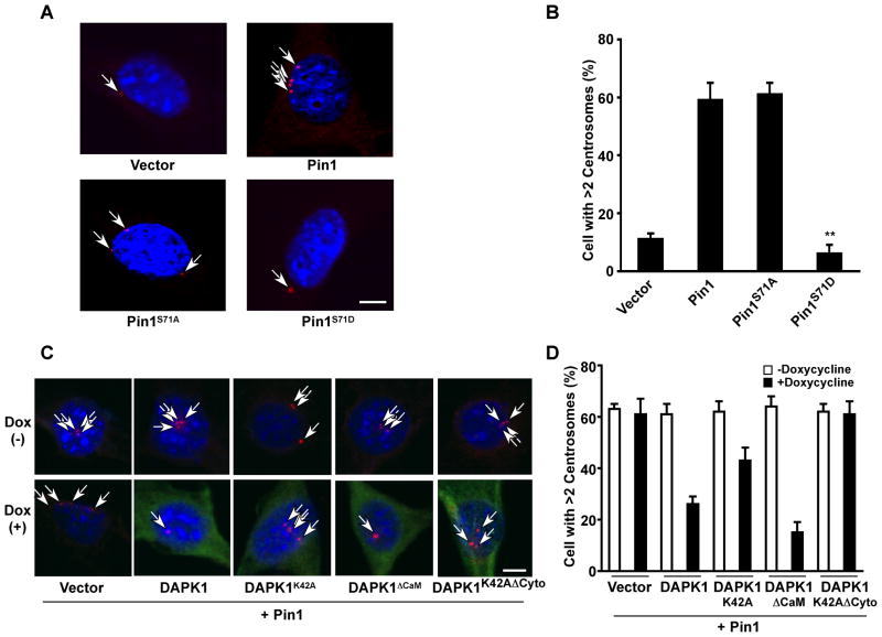
Figure 5. DAPK1 suppresses Pin1-induced centrosome amplification.
(A and B) S71D, but not S71A abolishes the ability of Pin1 to induce centrosome amplification. NIH3T3 cells stably expressing Pin1, its Ser71 mutants or control were arrested at the G1/S boundary by aphidicolin. Cells were stained with anti-γ-tubulin antibody (red) and DAPI (blue). Bar, 10 μm (A). Cells containing >2 centrosomes were scored in 300 transfected cells (B). Results shown are mean ± SEM, n = 3. **, p <0.001.
(C and D) DAPK1 inhibits Pin1-induced centrosome amplification. Stable cell lines were arrested at the G1/S boundary by aphidicolin in the presence or absence of doxycycline. Cells were stained with anti-γ-tubulin antibody (red) and DAPI (blue). Bar, 10 μm (C). Cells containing >2 centrosomes were scored in 300 transfected cells (D). Results shown are mean ± SEM, n = 3.

