
Figure 2.
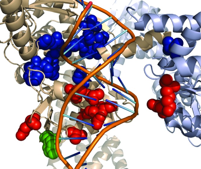
Location of amino acids that confer resistance to 1. The E. coli DNA gyrase structure from ref (22) (PDB 3NUH). The cartoon shows the GyrA subunit in beige, the GyrB subunit in gray, and the DNA duplex with the backbone and bases represented by lines. The DNA duplex was modeled to the structure by alignment of the GyrA subunit to the structure from ref (10) (PDB 2XCS). The atoms of several different amino acid side chains are displayed as colored spheres. The active-site tyrosine is green, amino acids that were identified in mutants that are resistant to quinolones are red, and amino acids that confer resistance to 1 are blue. The image was generated using PyMol.23