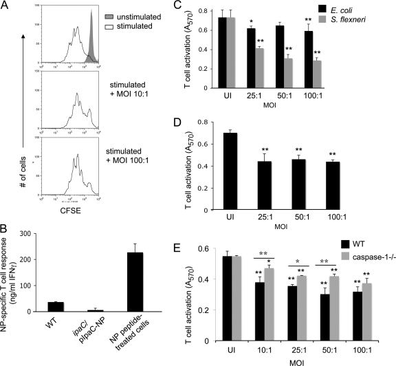
Fig. 4.
Antigens delivered into the cytoplasm of Shigella-infected cells fail to simulate antigen-specific CD8+ T cells. (A) CFSE-labeled naïve T cells were infected with S. flexneri at the indicated MOIs for 3 h and subsequently stimulated for 3 days by anti-CD3ε/anti-CD28 ligation in the presence of penicillin and streptomycin. Cells were gated on live cells. (B) Infected or NP118-126-coated J774 cells were incubated with NP118-126-specific CD8+ T cells and irradiated syngeneic spleen cells. Following 12 h of incubation, the amounts of IFN-γ present in the culture supernatants were determined by ELISA. (C to E) C57BL/6 BMM (C and E), 1308.1 epithelial cells (D), or caspase 1−/− BMM (E) were infected with S. flexneri or E. coli at the indicated MOIs and subsequently incubated with LFn-Ova257-264 and PA for 1 h. B3Z T cells were added, and at 8 h postinfection, B3Z T-cell activation was determined as described in Materials and Methods. Values represent the means of measurements for triplicate samples of a representative experiment, and error bars indicate SD. UI, uninfected. Data are representative of 3 (A), 2 (B), or at least 5 (C to E) experiments. For panels C to E, ANOVA and Bonferroni simultaneous tests were used for statistical analysis. Unless indicated otherwise, significance is for comparisons to uninfected congenic control cells (*, P < 0.05; **, P < 0.01.) For panel E, values for WT-infected cells were significantly different (P < 0.01) from those for uninfected WT cells at all MOIs tested. Values for caspase-1−/− infected cells were significantly different at all MOIs tested from those for uninfected caspase-1−/− cells. Differences between WT and caspase-1−/− cells were significant at MOIs from 10:1 to 50:1.

