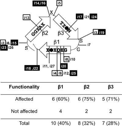
Fig. 2.
Localization of the insertion mutants in the coil model of TibA. One repeat of the consensus sequence is predicted to form three β-strands corresponding to one coil of a β-helix. A filled circle represents an isoleucine, a leucine, or a valine residue, while “X” represents any residue. Insertion mutants are positioned in this model. Mutants with a defect in autoaggregation (□) and mutants with a defect in adhesion (▪) are highlighted. The number as well as the percentage of insertions in each strand are shown below.

