
Fig. 3.
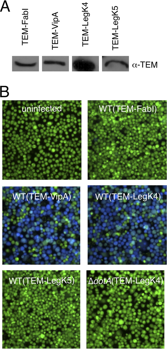
Dot/Icm-dependent translocation of LegK4 into J774 cells. (A) Western blot analysis of TEM fusion expression detected with an α-TEM antibody. (B) J774 cells were infected with L. pneumophila wild-type or dotA mutant strains harboring TEM-FabI, TEM-VipA, TEM-LegK4, and TEM-LegK5 expression plasmids at an MOI of 50. Infected cells were loaded with CCF4/AM, and translocation was determined by a comparison of cleaved to uncleaved CCF4 that gives blue and green fluorescence, respectively. Images were obtained by using epifluorescence microscopy on individual assay wells.