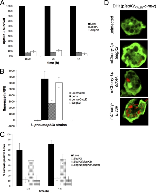
Fig. 6.
LegK2 is required for ER recruitment on LCV. (A) Uptake and survival ability of legK2 mutant strain. A. castellanii cells were infected at an MOI of 10 with wild-type, dotA mutant, and legK2 mutant strains. After different periods of contact with L. pneumophila, monolayers were treated for 1 h with gentamicin to kill adherent bacteria and disrupted with 0.04% Triton X-100. Viable intracellular bacteria were diluted and plated onto BCYE agar plates for colony enumeration. The results are expressed as a relative value (%) compared to a control invasion experiment with the wild-type strain. These data are representative of three independent experiments performed in triplicate; error bars represent the standard deviations. (B) Bacterial uptake assay. A. castellanii cells were infected with fluorescein-labeled Legionella at an MOI of 20, in the presence of cytochalasin D when indicated (+ CytoD). After 30 min of incubation, the medium was replaced by trypan blue solution to quench the fluorescence of noninternalized bacteria. The fluorescence of internalized bacteria was measured using an excitation of 485 nm and an emission of 530 nm. Fluorescence data were corrected for differences in labeling efficiency between the tested strains. Labeling efficiencies between strains varied by ca. 10%. (C) Recruitment of calnexin-GFP. Fifty Legionella containing vacuoles were scored from each sample by confocal laser scanning micrographs of calnexin-GFP-labeled D. discoideum AX3 infected at an MOI of 100 with mCherry-labeled L. pneumophila. Calnexin-positive vacuoles were numbered for amoeba cells infected by the wild-type L. pneumophila Lens strain, its derivative dotA and legK2 mutant strains, and the complemented legK2(plegK2) and legK2(plegK2K112M) mutant strains. The data are representative of three independent experiments; each error bar represents the standard deviation. (D) Localization of ectopically produced LegK2(K112M)-c-Myc in D. discoideum cells during infection. Confocal laser scanning micrographs of DH1 cells expressing legK2(K112M)-c-myc, either uninfected or infected with mCherry-labeled L. pneumophila legK2 mutant and dotA mutant strains or XL1-Blue E. coli. LegK2(K112M)-c-Myc was detected by an α-c-Myc antibody. The experiments were reproduced twice with similar results.

