
Fig. 3.
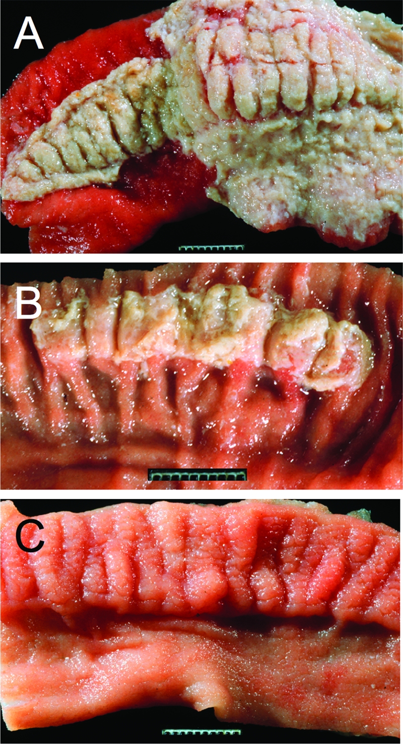
T3SS-1 and T3SS-2 contribute to intestinal inflammation in the calf gastroenteritis model. (A to C) Gross pathological appearance of the luminal surface of the terminal ileum collected 2 days after oral infection of calves with the S. Typhimurium wild-type (A), a T3SS-2-deficient mutant (B), or a T3SS-1-deficient mutant (C). Compared to the severe lesions caused by infection with the wild type (severe acute fibrinopurulent necrotizing enteritis with segmental or continuous pseudomembrane formation [A]), intestinal inflammation is reduced in calves infected with a T3SS-2-deficient mutant (moderate to marked subacute fibrinopurulent enteritis often confined to Peyer's patches [B]) and absent in calves infected with a T3SS-1-deficient mutant (normal Peyer's patch and ileal mucosa [C]). (Reprinted from reference 86.)