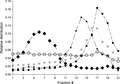
Fig. 4.
Subcellular localization of GS-9191-derived metabolites in SiHa and HEL cells. SiHa (○) and HEL (▿) cells were incubated with 14C-labeled GS-9191 (10 μM; 0.521 μCi/ml) for 4 h. The cells were lysed, and the organelles were separated on a Percoll gradient as described in Materials and Methods. Shown are the lysosomal marker hexosaminidase (■), the mitochondrial marker succinate dehydrogenase (▴), and the endoplasmic reticulum marker NADPH cytochrome c reductase ( ).
).

