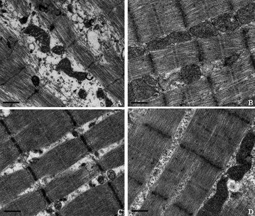
Fig. 6.
Representative electron photomicrographs of skeletal muscle from Himalayan marmots treated with metacavir (PNA), zidovudine (AZT), or water for 90 days. (A) AZT; (B) PNA at 100 mg/kg/day; (C) PNA at 50 mg/kg/day; (D) control. Bars show 1 μm. Ultrastructural features include registered sarcomeres with thick and thin filaments and mitochondria. Swelling of mitochondria, giant mitochondria, disruption of mitochondrial membranes, and sarcomere disruptions with Z-line misalignment are observed in the AZT-treated group. Conversely, no obvious mitochondrial morphological changes are observed in the PNA-treated and control groups. Original magnification, ×18,500. In each sample, more than 10 fields were examined under low or high magnification.

