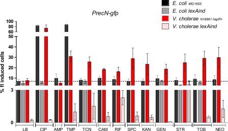
Fig. 1.
SOS induction by subinhibitory concentrations of antibiotics in E. coli and V. cholerae. Bacteria were grown overnight in MH supplemented with sub-MICs of specified antibiotics as follows (final concentrations [μg ml−1] of drug for organism [E. coli, V. cholerae]): ciprofloxacin (CIP), 0.05, 0.005; ampicillin (AMP) and rifampin (RIF), 0.05, 0.01; trimethoprim (TMP), 0.05, 0.005; tetracycline (TCN), 0.15, 0.015; chloramphenicol (CAM), 0.15, 0.005; spectinomycin (SPC), kanamycin (KAN), and streptomycin (STR), 0.2, 0.05; gentamicin (GEN), tobramycin (TOB), and neomycin (NEO), 0.1, 0.01. The samples were washed in PBS, and the fluorescence was measured using the FACSCalibur device (1). The percentage of fluorescent cells is represented on the y axis. Each measurement was reproduced at least four times. In order to test whether the means of two groups were different, we first tested the equality of their variance with a Fisher test. When the variances of two groups were found to be significantly different, an unequal-variance t test was performed. Otherwise, a simple t test was performed. The chosen significance threshold was 0.05 for all tests.

