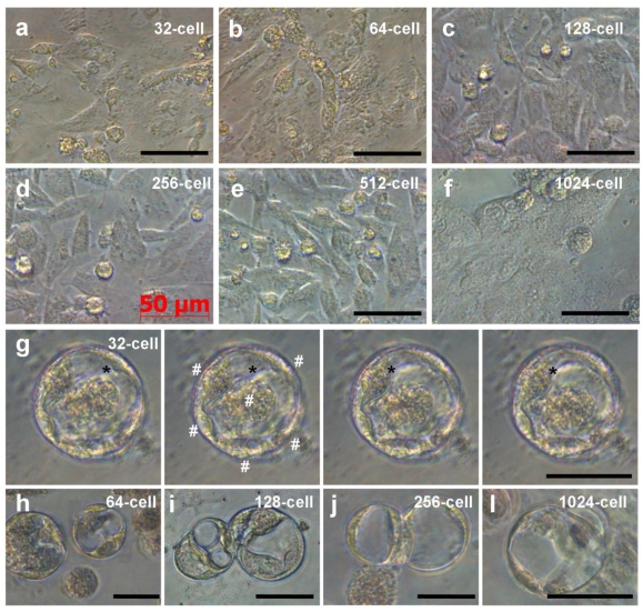
Fig 4.
Embryonic cells of different stages during 6 days of culture. (a-f) ES-like cells from embryos at indicated stages. (g) Serial micrographs of YSL cell, showing the motility of the cytoplasm (asterisks) residing in the center and multiple nuclei (#) at the periphery. (h-l) YSL cells in cultures from embryos at stages indicated. Scale bars µm.

