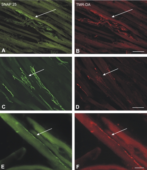
Figure 2.
Combined immunofluorescence of the tracer TMR-DA (red) and SNAP-25 (green) identifying en plaque (A, B), en grappe (E, F), and palisade endings (C, D) in the medial rectus muscle of the monkey in case 1. Each pair of neighboring photographs shows the same section with different fluorescence filters. (A, B, E, F) Micrographs were obtained with a fluorescence microscope (DMRB; Leica, Bensheim, Germany) and (C, D) with a confocal microscope (TCS SP; Leica). Scale bar: (A–D) 50 μm; (E, F) 100 μm.

