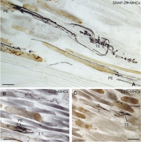
Figure 4.
Bright-field immunoperoxidase staining for MHCs (brown) combined with either SNAP-25 (black) or CTB (black). (A) A SNAP-25-positive palisade ending (PE) (black) contacts an MHCs-positive MIF in the top left corner; its axon is seen to be continuous with a tendon ending (TE) in the superior oblique tendon (T). (B, C) CTB labeled palisade endings (black) and labeled tendon ending (black) in the MR muscle, after tracer injection. (case 2). (All images obtained with a DMRB microscope; Leica, Bensheim, Germany). Scale bar, 50 μm.

