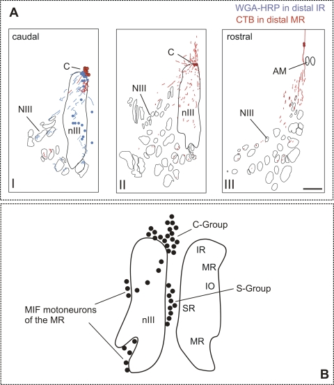
Figure 5.
(A) Reconstruction of caudorostrally arranged transverse brain stem sections demonstrating motoneurons (dots) in the C-group (C) that have been tracer-labeled by injections into the myotendinous junction of the medial rectus (MR; red) and inferior rectus (IR, blue). The respective tracer-labeled axons of both populations travel through the oculomotor nucleus (nIII) and would be labeled by injections into nIII. Scale bar, 500 μm. NIII, oculomotor nerve; AM, anteromedian nucleus. (B) A summary diagram of the total population of neurons around and within the oculomotor nucleus (nIII) after injection into the myotendinous junction of all eye muscles, including the C-group and S-group.21 Present results suggest that they contain both MIF-motoneurons and palisade ending somata.

