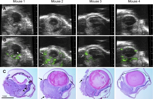
Figure 4.
Samples of uveal melanomas imaged in four mice with 2D ultrasound before (A) and after (B) contrast enhancement and correlated histologic sections (C, hematoxylin and eosin, ×40). Funnel-shaped retinal detachment in mouse 1 (arrowheads) and extraocular extension of tumor in mouse 2 and mouse 3 was observed with enhancement (arrows).

