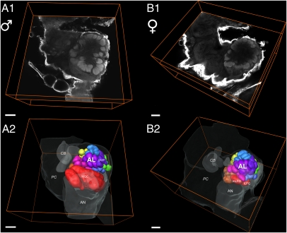
Fig. 3.
Example of the right hemisphere of the Manduca brain with focus on the AL and its innervation by antennal nerve fibers (AN). (A) Confocal micrographs extracting one single optical orthogonal slice from the 3D dataset of one male (A1) and one female AL (A2). (B) 3D reconstructions of the AL of both sexes depicting a ventral view of reconstructed glomeruli (B1: males, n = 68; B2: females, n = 70 ± 1) corresponding to optical slices in A. ALs display typical moth glomerular architecture: the striking sex-specific glomeruli, the male-specific MGC, and the medial large (mLFG) and lateral large female glomeruli (lLFG) are situated at the entrance of the antennal nerve and are illustrated in red. Brain outlines of adjacent neuropil areas serve as orientation guidelines. CB, central body; PC, protocerebrum; OL, optic lobe; AN, antennal nerve; glom, glomerulus. (Scale bars: 100 μm.)

