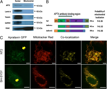
Fig. 2.
Targeting of aprataxin to mitochondria. (A) Western blotting of purified extracts confirms mitochondrial localization of aprataxin. Membrane was probed with antiaprataxin antibody (ab31841), which detected an ∼40-kDa band in both nuclear and mitochondrial extracts. Lamin b and GAPDH/Tubulin antibodies were used to detect nuclear or cytoplasmic contamination of mitochondrial extracts, respectively. The mitochondrial protein VDAC was used to confirm mitochondrial enrichment of the mitochondrial fraction; 20 μg protein were loaded in each lane. Blot is representative of four separate experiments. (B) Full-length protein and two relevant isoforms of aprataxin. Isoform b has a distinct 14-aa N-terminal stretch that harbors the putative mitochondrial targeting sequence (MTS). The probability of mitochondrial localization was estimated using the mitochondrial prediction software MitoProt. FHA, fork head-associated; NLS, nuclear localization signal; HIT, histidine-triad; ZNF, zinc finger. Antigenic region for the aprataxin antibody (amino acids 1–177 of isoform a) is denoted. (C) C-terminal GFP tagged isoform b aprataxin (Aprataxin GFP) localizes to the mitochondria. The location of the fusion protein was compared with the Mitotracker Red mitochondrial marker. Additional details are in Fig. 1. The nucleus is indicated with a yellow arrow. (Scale bars: SH-SY5Y, 20 μm; NT2, 10 μm.)

